Pulmonology: Respiratory & Lung Care
417.347.8315
Breathe More Easily with Freeman Lung Institute
It's important to get the right care for pulmonary conditions, and that’s why the lung care specialists at Freeman Lung Institute provides clinical expertise, state-of-the-art diagnostic services and access to the latest research-based medical advancements, surgical procedures and rehabilitation therapies. Our advanced lung disease experts offer inpatient and outpatient treatment for a full range of pulmonary issues to get you the care you need. They help you understand and manage your condition.
Our respiratory specialists provide a continuum of care by collaborating with colleagues in other specialties as needed, including radiation oncology, cardiology and cardiovascular surgery, radiology, pathology, hematology, infectious diseases and more.
Common signs of lung problems include:
- Trouble breathing
- Shortness of breath
- Feeling like you're not getting enough air
- Decreased ability to exercise
- A cough that won't go away
- Coughing up blood or mucus
- Pain or discomfort when breathing in or out
Our lung doctors diagnose and treat a variety of lung conditions:
- Chronic obstructive pulmonary disease (COPD)
- Interstitial lung disease
- Asthma
- General pulmonology
- Lung cancer
- Pulmonary rehabilitation
- Pulmonary vascular disease
- Sarcoidosis
- Abnormal buildup of fluid in the lungs (pulmonary edema)
- Lung infection (pneumonia)
- Collapse of part or all of the lung (pneumothorax or atelectasis)
- Swelling and inflammation in the main passages (bronchial tubes) that carry air to the lungs (bronchitis)
- And more
LUNG CARE SERVICE
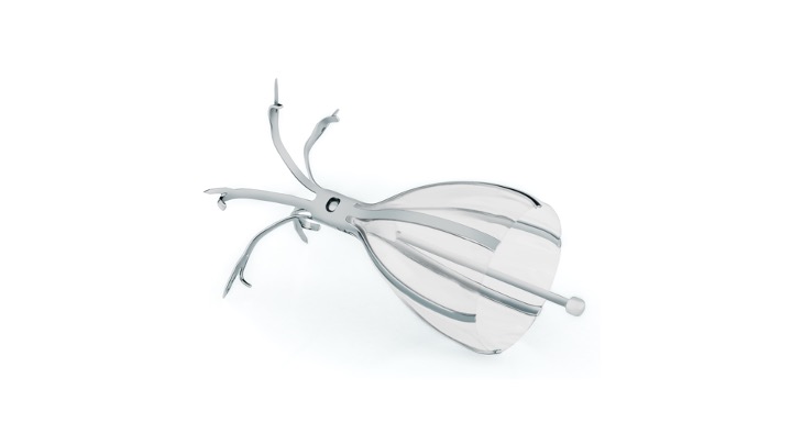
Spiration Valve System
Freeman is the first and only health system in the area to offer the Spiration Valve System (SVS), which can help alleviate your emphysema symptoms. This safe and effective minimally invasive treatment option is proven to improve lung function, reduce shortness of breath and restore quality of life in those suffering from severe emphysema.
How it works
Air breathed in is redirected away from the diseased part of the lung to the healthier parts. Air and fluids trapped in the diseased part of the lung can then escape. Surgery is not required to place the valve in the airways because the doctor uses a bronchoscope to place the spiration valve. Valves are placed inside the airways using a catheter. The valve then expands and contracts with breathing. More information on SVS can be found at svs.olympusamerica.com.
LUNG CARE SERVICE
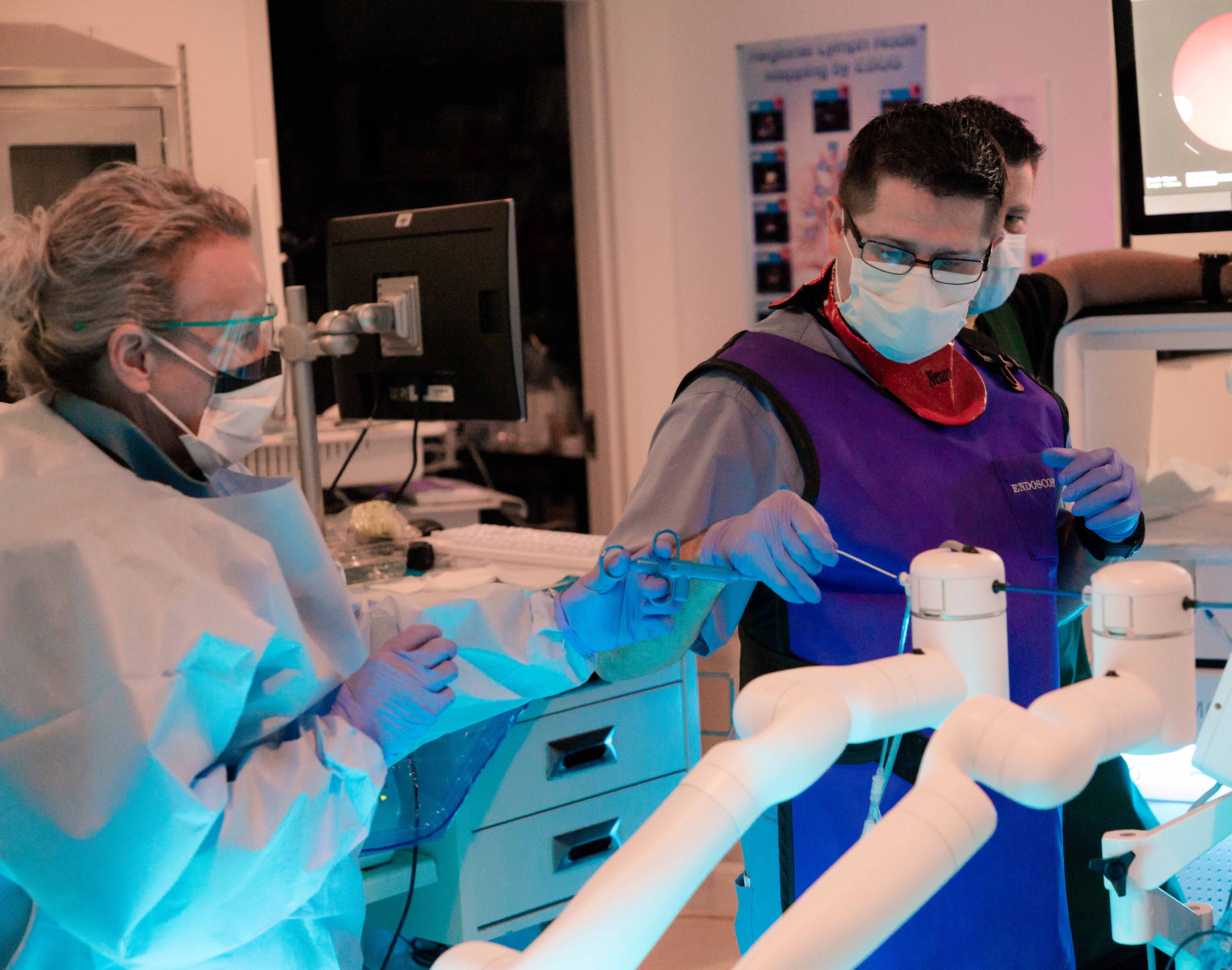
Freeman Lung Institute pulmonologists use the transformative MONARCH® robotic bronchoscopy platform, a unique technology targeting lung cancer and a better way to see inside a patient’s lungs with a precision tool (pinpoint precision).
The MONARCH robotic-assisted bronchoscope is a game-changer for patients seeking a diagnosis of a nodule in their lungs. Its novel telescoping design enables physicians to reach farther into the lungs and visualize what is being biopsied, all with impressive precision. Each component of the bronchoscope can be independently articulated, advanced, retracted and positionally locked, enabling physicians to have greater control and maneuverability deep in the lung, where most small nodules are found.
Lung cancer is the leading cause of cancer-related death in the United States, and early diagnosis is key to successful treatment. The problem is symptoms don’t often appear in the early stages of the disease, and once symptoms appear, it’s often a more advanced stage of a cancer that has either grown to a large size or spread throughout the body.
The combination of better reach, visualization and control provides our pulmonologists an advanced method for diagnosing early-stage lung cancer when it’s most treatable. Added benefits include no incisions made, fast recovery time (patients usually go home the same day), saves time and reduces stress by eliminating the need to make multiple trips to the hospital, and any “positive” nodules that are discovered can be treated immediately.
Freeman is the first hospital in Missouri, Southeast Kansas and Northeast Oklahoma to invest in the MONARCH® Platform by Auris Health, Inc. that ultimately combines precision robotics, software, data science and endoscopy (the use of small cameras).
For more information about MONARCH, pulmonology services or to make an appointment, call 417.347.8315.
LUNG CARE SERVICE
Inspire® therapy
Freeman is the first and only health system within a 70-mile radius of Joplin to offer Inspire® therapy, a new treatment option for patients with obstructive sleep apnea. This innovative solution can offer relief to sleep apnea sufferers who cannot tolerate continuous positive airway pressure (CPAP) therapy.
Inspire is the only FDA-approved OSA treatment that works inside your body to treat the root cause of sleep apnea. Inspire works inside the body with a patient’s natural breathing process to treat sleep apnea. Mild stimulation opens the airway during sleep, enabling oxygen to flow naturally. The patient uses a small handheld remote to turn Inspire on before bed and off when they wake up.
To learn more about Inspire, visit freemanhealth.com/ent.

LUNG CARE SERVICE
Endobronchial Ultrasound (EBUS) Bronchoscopy
Endobronchial ultrasound (EBUS) bronchoscopy is an interventional procedure using ultrasound technology in combination with bronchoscopy to visualize the airway walls and surrounding structures including inflammation, infections or cancer.
EBUS enables Freeman pulmonologists to locate hard-to-reach tumors and small cell lung cancer. It can also be used to take a biopsy from tissue in the lungs or from the surrounding lymph nodes in the chest. Those samples can be used for diagnosing and staging lung cancer, detecting infections and identifying inflammatory diseases that affect the lungs, such as sarcoidosis or cancers like lymphoma.
EBUS can be performed on an outpatient basis instead of requiring a more complex procedure or surgery. Additionally, it may reduce the number of required procedures as well as the number of complications that can arise from other diagnostic methods.
Performed by pulmonologists at Freeman Lung Institute, EBUS bronchoscopy uses a flexible tube that goes through the mouth and into the windpipe and lungs. The EBUS scope has a video camera, with an ultrasound probe attached, to create local images of the lungs and nearby lymph nodes to accurately locate and evaluate areas seen on x-rays or scans that require a closer look.
During a conventional diagnostic procedure, surgery known as mediastinoscopy, is performed to provide access to the chest. A small incision is made in the neck just above the breastbone or next to the breastbone. Next, a thin scope, called a mediastinoscope, is inserted through the opening to provide access to the lungs and surrounding lymph nodes. Tissue or fluid is then collected via biopsy. An endobronchial ultrasound is much less invasive.
Benefits of EBUS include:
- Real-time imaging of the surface of the airways, blood vessels, lungs and lymph nodes
- Improved images enable the physician to easily view difficult-to-reach areas and to access more and smaller lymph nodes for biopsy
- The accuracy and speed of the EBUS procedure lends itself to rapid onsite evaluation
- EBUS can easily and safely be performed under conscious sedation within reasonable time
- Patients recover quickly and can usually go home the same day
Lung service
Pulmonary Function Testing (PFT)
PFT is a non-invasive, painless test demonstrating how well the lungs are working. PFT measures lung volume, capacity, rates of flow and gas exchange. Testing is completed by a plethysmograph where patients sit or stand in an air-tight box, which looks similar to a small telephone booth. Not only does PFT save patient time, peace of mind and money, but PFT also provides immediate results and instant treatment, if needed.
Who benefits from PFT?
While testing can be completed during a routine physical, most of the time it is for patients with a lung condition. PFT assists healthcare providers to determine diagnoses, such as:
- Allergies
- Respiratory infections
- Chronic lung conditions, such as asthma, bronchiectasis, emphysema or chronic bronchitis
- Asbestosis
- Sarcoidosis
- Restrictive Airway problems from scoliosis, tumors, inflammations or scarring of the chest wall
LUNG CARE SERVICE
Ultrasound-Guided Thoracentesis for Pleural Effusions
An ultrasound-guided thoracentesis is a procedure in which a needle is inserted through your chest wall into your lung cavity to remove or collect fluid accumulation (called a pleural effusion).
Doctors use thoracentesis to:
- relieve pressure on the lungs
- treat symptoms such as shortness of breath and pain
- determine the cause of excess fluid in the pleural space
How is the procedure performed?
A chest x-ray may be performed before a thoracentesis.
This procedure is often done on an outpatient basis. However, some patients may require admission following the procedure. Ask your doctor if you will need to be admitted.
The doctor or nurse will position you on the edge of a chair or bed with your head and arms resting on an examining table.
They will sterilize the area of your body where the needle is to be inserted and cover it with a surgical drape.
Your doctor will numb the area with a local anesthetic. This may briefly burn or sting before the area becomes numb.
The doctor inserts the needle through the skin between two ribs on your back. When the needle reaches the pleural space between the chest wall and lung, the doctor removes the pleural fluid through a syringe or suction device.
Thoracentesis usually takes about 15 minutes.
At the end of the procedure, the doctor will remove the needle and apply pressure to stop any bleeding. They will cover the opening in the skin with a dressing. No sutures are necessary.
If necessary, you may have a chest x-ray after thoracentesis to detect any complications.
Generally, once the procedure is completed, the patient is further educated about discharge instructions and is released to go home (as appropriate).
What does the equipment look like?
In this procedure, ultrasound, CT, or x-ray equipment may be used to guide a needle into the fluid within the pleural space. Thoracentesis is typically performed with ultrasound guidance. Occasionally, CT-guidance will be used.
Ultrasound machines consist of a computer console, video monitor and an attached transducer. The transducer is a small hand-held device that resembles a microphone. Some exams may use different transducers (with different capabilities) during a single exam. The transducer sends out inaudible, high-frequency sound waves into the body and listens for the returning echoes. The same principles apply to sonar used by boats and submarines.
The technologist applies a small amount of gel to the area under examination and places the transducer there. The gel enables sound waves to travel back and forth between the transducer and the area under examination. The ultrasound image is immediately visible on a video monitor. The computer creates the image based on the loudness (amplitude), pitch (frequency), and time it takes for the ultrasound signal to return to the transducer. It also considers what type of body structure and/or tissue the sound is traveling through.
The CT scanner is typically a large, donut-shaped machine with a short tunnel in the center. You will lie on a narrow table that slides in and out of this short tunnel. Rotating around you, the x-ray tube and electronic x-ray detectors are located opposite each other in a ring, called a gantry. The computer workstation that processes the imaging information is in a separate control room. This is where the technologist operates the scanner and monitors your exam in direct visual contact. The technologist will be able to hear and talk to you using a speaker and microphone.
A thoracentesis needle is generally several inches long and the barrel is about as wide as a large paper clip. The needle is hollow so fluid can be aspirated (drawn by suction) through it. In some instances, a small tube is advanced over the needle, and the fluid is removed through the tube after removing the needle.
What are the benefits vs. risks?
Benefits
Thoracentesis is generally a safe procedure
No surgical incision is necessary
No scheduled appointment is necessary
Risks
Any procedure that penetrates the skin carries a risk of infection. The chance of infection requiring antibiotic treatment appears to be less than one in 1,000.
Freeman Health
Lung Institute
Freeman Lung Institute features best-in-class pulmonary function testing (PFT), which is painless and offers immediate results and instant treatment right in the physician’s offices. The clinic, the first and only in the area, gives patients access to screening, diagnosis, treatment and support of life-threatening lung diseases in one convenient location.
Freeman Lung Institute provides comprehensive care for a wide range of pulmonary conditions including:
- Lung cancer
- Chronic Obstructive Pulmonary Disease (COPD)
- Emphysema
- Asthma
- Pulmonary hypertension
